Introduction
Dr. Yaohui (Gloria) Xu, MD, PhD, is a Professor and Section Chief of Mohs Dermatologic Surgery in the Department of Dermatology at the University of Wisconsin-Madison in the School of Medicine and Public Health. She is board certified in dermatology as well as Mohs Dermatologic Surgery and the Director of Micrographic Surgery and Dermatologic Oncology Fellowship. Her educational interests include training medical students, dermatology residents, fellows, and general practitioners. Since 2016, Dr. Xu has been a panel member for the national guideline-establishing group, the National Comprehensive Cancer Network (NCCN) Skin Cancer Excluding Melanoma Guideline. Her recent publication, “Prevalence of poor outcomes in cutaneous squamous cell carcinoma by AJCC and BWH tumor stages: A systematic review and meta-analysis,” was published in January 2025 in the Journal of the American Academy of Dermatology.
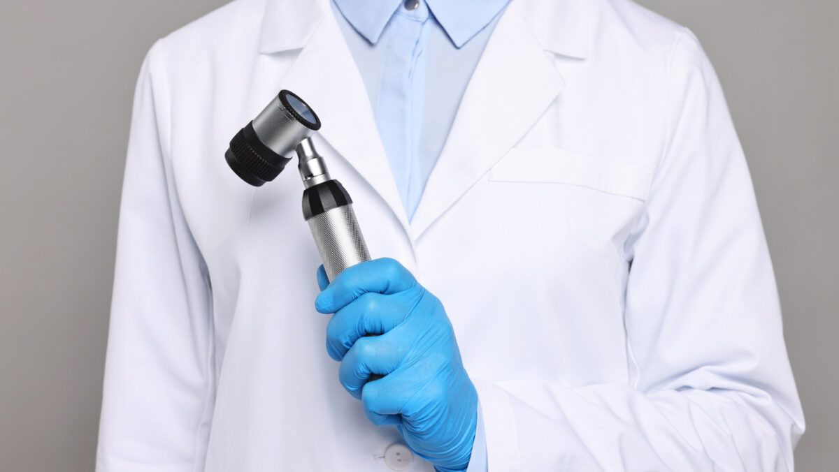
What are Mohs Surgery and Wide Local Excision?
Mohs surgery and wide local excision are surgical procedures clinicians use to remove skin cancers. When performing wide local excision or Mohs surgery, surgeons and dermatologists already know there is cancer because a skin cancer diagnosis has been made. Either wide local excision or Mohs surgery is used for treatment. The patient diagnosed has a skin cancer that needs removal.
The term “wide local excision” describes how the surgeon performs the surgical technique. After wide local excision, what’s next determines how the pathologist processes the tissue. Using wide local excision, the cure rate for nonmelanoma skin cancer is approximately 95%. The cure rate is good; the technique is practical and balanced between the resources required and the results produced.
Mohs surgery is another method for removing skin cancer. It uses a sequential, repeatable technique to remove a small amount of tissue. Although this blog is designed to provide information on wide local excision, readers can find a more in-depth explanation of Mohs here. In addition, a history of Mohs surgery is available here.
What are the Major Differences between the Procedures?
The Amount of Tissue Removed
The amount of tissue removed is the fundamental difference between these techniques. Mohs surgery and wide local excision are also very different in terms of the pathologic margins cut and the amount of tissue that is checked.
For Mohs surgery, the Mohs surgeon can cut smaller and shallower portions of tissue, essentially cutting tissue more conservatively and sparing the healthy tissue. In wide local excision, the dermatologist or surgeon has to cut bigger and deeper portions of tissue with a pre-determined margin.
Using wide local excision for ordinary basal cell carcinoma, the dermatologist will cut at 4 mm margin. The margin is the circle that is 4 mm bigger, or the peripheral margin, around the area. For squamous cell carcinoma, the average peripheral margin is usually 5 mm. Why do we use 4 and 5 mm? We use these sizes for basal and squamous cell carcinoma because the guidelines say to do so. The guidelines provide evidence-based information, and when the evidence is lacking, expert consensus is used.

For melanoma, a dermatologist would cut even bigger portions of tissue if they were using the wide local excision technique. In Stage I melanoma, the guidelines indicate a dermatologist should cut 1 cm of the peripheral margin. This means that a dermatologist would draw a circle 1 cm out from around the lesion. So right away, even if you have a pinky-sized lesion, if you draw 1 cm circle around that spot, it’s more than a quarter (coin) in size.
For Mohs surgery, a dermatologist typically cuts 1-2 mm around the border of the tumor. Instead of 4-5 mm like wide local excision, a dermatologist cuts 1-2 mm with Mohs, which is much smaller and more conservative at the first round of Mohs surgery. The reason clinicians can cut more conservatively with Mohs surgery is that the margins are checked differently.
To put these numbers into perspective, 1 cm is about the width of a pencil eraser and 1 mm is about the width of the sharpened point of a pencil used for writing. One cm is also equivalent to 10 mm. In other words, these measurements are tiny.
Tissue Processing Using Bread Loafing Method in Wide Local Excision
Prior to having and using Mohs surgery, the traditional way to check the specimen and its margins during wide local excision was done using a method or process called “Bread Loafing.” The term Bread Loafing describes the way that a pathology technician cuts skin cancer tissue specimens into sections and then checks the tissue.
For example, when cutting bread, you slice across in cross sections like left-to-right, not top-to-bottom. The pathology technician will cut the tissue using Bread Loafing for tissue processing. They cut from one end to the other end of the specimen. These cross cuts from side to side produce thin cross sections which can be placed down on glass slides. As an example, this would be similar to cutting bread from a loaf for a square-shaped, traditional sandwich, not a long length sub bun.

Although they cut across from one end to the other using the Bread Loafing technique, like cutting sandwich bread slices, the technician does not cut continuously. Instead, the cuts made by the technician may skip 2-4 mm of tissue and then keep going.
In other words, one might cut a slice of bread from one end and then skip all the bread in the middle, before cutting another slice of bread from the opposite end of the loaf. Therefore, you may have a (2-4 mm) gap between each cross-section of your bread. Technicians do not cut continuously. They skip around.
The technician skips around in the tissue because if all the sections were cut throughout the entire lesion, there would be far too many sections for the pathologist to read. It would result in too many slides, too many materials, and too much of the pathologist’s time, and most of the time, results determine nothing new. It’s not practical at all and not useful.
If you do the calculations involved with this process, it proves that Bread Loafing in wide local excision only visualizes a small percentage of tumor margins. As an example, using Bread Loafing, with the technician skipping 2-4 mm of the specimen between each section cut, only 1% of the tumor margin is actually examined in wide local excision. Therefore, Bread Loafing used in wide local excision has its inherent limitations if you want to examine tissue margins.
However, Bread Loafing is important for diagnosis. If we have a rash or tumor on the skin, the clinician may not know exactly what it is. In this case, bread loafing is important to sample part of the lesion. The lesion can be cut in the middle, or where it is biologically important to the rash or tumor process. Then, the clinician can see the disease process and make a diagnosis.
The Margins (or Edges where the Tumor Contacts Normal Healthy Tissue)
When it comes to margins, the margins for Mohs surgery are different than those for wide local excision, starting with the type of cuts made by Mohs surgeons. With Mohs cutting, it is not a straight, vertical, 90-degree cutting. Instead, it is cut using a bevel of the blade to produce a specimen cut in the shape of a pie.
The Mohs surgeon cuts a piece of the skin tissue out, and it looks like a round, oval-shaped pie. The top is a (skin/lesion) crust, and the bottom is blood and fatty tissue. Instead of cutting like Bread Loafing, the technician is trained to smash the pie on a flat surface and then freeze it that way.
When the technician smashes or flattens the pie, the goal is to make the “peripheral margin,” or the edge of the pie, and the base of the pie to be on the same level or in the same plane.
The smashed pie is then cut horizontally–from the bottom of the pie and then up through the pie crust at the top. The cut will capture peripheral and deep margins on the same cut.
That’s what is good about the Mohs method—the ability to check margins on one slide. Depending on the size of the tumor, peripheral and deep margins are captured together. Theoretically, if the tumor is small enough, you can put everything on one glass slide.
That is why we can say accurately that we check margins at 100% with Mohs surgery. The smashed pie specimen provides a view of the peripheral and deep tissue fields at the same time. With a skilled technician, they should not miss anything – Mohs surgeons are seeing 100% tumor margins. If cancer is positive on the Mohs margin, the Mohs surgeon will repeat the process and remove the residual skin cancer either wider (if the peripheral margin is positive with cancer) or deeper (if the deep margin is positive with cancer), specifically guided by microscopic reading.
With wide local excision, a doctor removes a lot more tissue overall to ensure the cancer is removed and checks 1% of the margins. Because the edges are not checked 100%, it is technically possible to miss some skin cancer nearby when using wide local excision. However, the cure rate using wide local excision is still decent, approximately 95%, due to the pre-determined margin being removed around the cancer.
How Does a Clinician Decide Which Method is Best?
Clinicians practicing in this area use very specific guidelines and standards. Mohs surgery is not for every skin cancer and it is a little more costly. In some cases, topical creams or “scrape and burn” method (technical term is called electrodesiccation and curettage) are likely to be more cost-effective for the patient. The scrape and burn method may work for a superficial basal cell cancer in a cosmetically unimportant location and a patient may not need Mohs surgery.
Other aspects of skin cancer and the patient play a role. For example, does the preliminary pathology report that the tumor is aggressive or not? The location of the skin cancer is also important, as is its size. A clinician may also factor in the age of the patient and, of course, a patient’s preferences.
Ultimately, the “Mohs Appropriate Use Criteria” assembled in collaboration with the American Academy of Dermatology, guides clinicians on the best practices. Using 270 different scenarios, expert judgement, and clinical practice, the document advises when it is reasonable to one procedure versus another.1
A clinician may consider the available medical information on the lesion (e.g., location, features), expert opinions, patient characteristics (e.g., immunocompromised, prior radiation), and the health care system to select one technique over another.1 Lastly, the referral doctors may also aid in the decision.
Conclusion
Mohs surgery and wide local excision are two different surgical procedures for removing nonmelanoma skin cancer. Major differences between these techniques include the amount of tissue removed and how the tissue is processed in the lab. WLE has cure rates about 95% and Mohs has cure rates 98-99% for primary nonmelanoma skin cancers. The patient and their health care providers make the final decision for treatment.
References
1. Ad Hoc Task Force; Connolly SM, Baker DR, Coldiron BM, et al. AAD/ACMS/ASDSA/ASMS 2012 appropriate use criteria for Mohs micrographic surgery: a report of the American Academy of Dermatology, American College of Mohs Surgery, American Society for Dermatologic Surgery Association, and the American Society for Mohs Surgery. J Am Acad Dermatol. 2012;67(4):531-50. doi: 10.1016/j.jaad.2012.06.009.
Recent Posts
Oct. 08, 2025
Regeneron Announces Approval of Cemiplimab-rwlc for Adjuvant Treatment of Cutaneous Squamous-Cell Carcinoma with a High Risk of Recurrence After Surgery and Radiation
Sep. 29, 2025
Preparing for Your First Oncology Visit: A Complete Guide for Skin Cancer Patients
Sep. 05, 2025
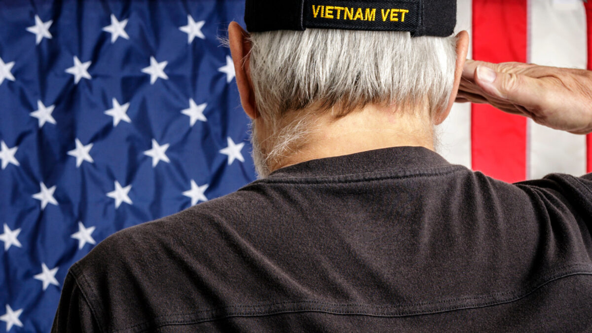
Skin Cancer in Aging Veterans: Unique Risks & Considerations
Aug. 25, 2025
The Genetics of Skin Cancer: Insights from Dr. Kenneth Tsai
Aug. 16, 2025