The widely used technique for removing nonmelanoma skin cancers, Mohs micrographic surgery, began in a small lab in Wisconsin during the 1930s. The technique’s name is not an acronym, but the name of its inventor, Dr. Frederic Mohs – a former surgeon in the Department of Surgery at Wisconsin General Hospital. During the initial use of Mohs surgery, there was some apprehension and misunderstanding about its medical science, as evidenced by a bid to revoke the medical license of its inventor. [Ross 2015] Presently, millions of patients have probably undergone Mohs surgery to remove suspicious skin lesions, both nonmelanomas and early invasive melanomas. In this article, we will briefly cover the history of the technique.
What exactly is Mohs micrographic surgery, and how is it performed?
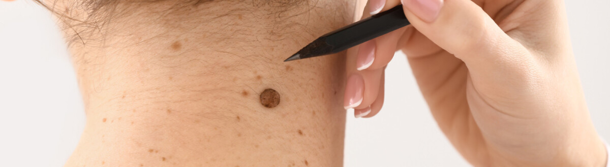
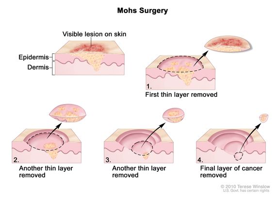
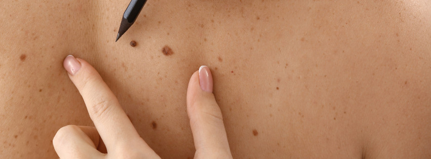
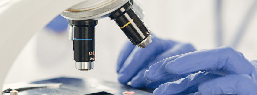
Mohs micrographic surgery is the gold standard method for removing nonmelanoma skin cancer, and it is used to remove some early-stage melanomas. Specifically, it is a serial technique that allows layers of a suspicious skin to be repetitively removed one at a time, as needed, during the procedure until no remaining areas look suspicious under a microscope. It allows the skin lesion to be removed with confidence, ensuring that the surrounding tissue looks healthy.
The procedure also minimizes the amount of tissue necessary for removal. This process is preferable to cutting off larger chunks of the skin, which can complicate wound healing and result in unnecessary scarring, without truly knowing whether all of the cancer was removed. Due to the repetitive remove-and-examine process, it may take several hours for a patient to undergo Mohs surgery completely.
The first ever rudimentary Mohs surgery was performed in the U.S. on June 30, 1936. The modern technique used globally is a refined version of that procedure.
Who was Dr. Frederic Mohs, M.D.?
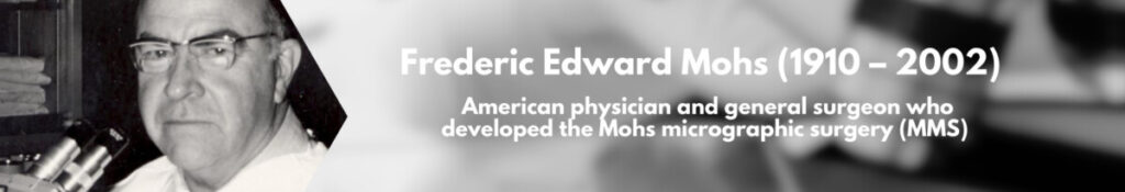
Fred Mohs (1910-2002) was a Wisconsin-born physician who received his undergraduate degree from the University of Wisconsin in Madison. He continued his education as a medical student and graduated from the School of Medicine and Public Health in 1934. Mohs trained as a general surgeon in Portland, Oregon, before returning to Madison to practice surgery and continue doing research. He is the pioneer and inventor of Mohs micrographic surgery.2
Originally, the Mohs family emigrated to Wisconsin from Prussia around 1848, and Fred Mohs’ father, Frederic Carl, was the first generation born in the U.S. As a family man, Fred Mohs married Mary Ellen, his companion-for-life, until she succumbed to a stroke in 1995. Together, they raised three children in Madison, where the Mohs family still lives. Fred Mohs died in 2002 at the age of 92 after being in mental decline. During his final years, his son described his demeanor as “nicer, more appreciative, and less intense.” 3
How did he invent the technique we refer to as “Mohs surgery?”
During college, Mohs worked as an undergraduate research assistant with animals in the Department of Zoology. As he gained more responsibility in the laboratory, he began to make observations about normal and cancer cells, the preservation of tissue, and the microscopic examination of tissues.
According to Dr. Juliet Aylward, M.D., Professor and Section Chief of Dermatologic Surgery at the University of Wisconsin School of Medicine and Public Health and Lead of the Mohs Oral History Project, “He wasn’t hooked on skin cancer to start with, he was a scientist first and a clinician second,” she said.
When Mohs returned to Madison to work as a professional, he set up a practice devoted to surgery. He also began experimenting with “chemosurgery” techniques, combining research solutions with surgery.
“He was looking for a model for cancer,” Aylward said. “The obvious model is skin because you can see it, touch it, and monitor it. Skin became a natural way for him to study cancer, which is really what he wanted to do.”
Aylward says that the Mohs surgery technique grew from the research that he began as an undergraduate and continued throughout his professional life. “He was devoted to treating skin cancer surgically; he had the surgical skills and the research background to study the skin as a model.”
Mohs needed to develop a method to capture a layer of skin without cutting directly into live tissue. The (incorrect) belief at the time was that cancer spreads through cutting.1 Removing it would, therefore, require a gentle yet thorough approach. Eventually, he developed a fixative paste that killed and preserved tissue so well that animal or human tissue looked comparably alive under the microscope, just as it had been.2
His early methods used a paste that softened the skin, allowing a layer to be easily scraped off without damaging it. The paste also included a fixative for the tissue, preserving it indefinitely.
Combining the pastes with the surgical removal allowed a layer of skin to be confidently removed from a patient and then analyzed under a microscope. Although this process is taken for granted today, that is merely due to Fred Mohs’s success in the lab and in the clinic.
What changes in Mohs surgery technique evolved over the years?
Shockingly, if a patient in the late 1930s had Mohs surgery performed, they did not receive any type of local anesthetic prior to skin removal.6 However, they were given a prescription for codeine or morphine, drugs that were becoming heavily controlled in the U.S. around that time.
Some were skeptical when anesthesia was introduced into the procedure in the 1940s. Now, local anesthesia is standard practice.
Without any anesthetic to numb the pain, early recipients of Mohs surgery felt their skin soften from acid and become fixed with a black paste. Later, they observed when it was scraped off by the surgeon. Understandably, the process was excruciating for early patients receiving Mohs surgery. The black paste is also no longer in use.
Modern Mohs surgery can sometimes take hours, but the original procedure took days to complete. The black fixative paste was applied after the top layer of skin debris was removed. Then, patients were seen again the following day for an additional layer of tissue removal. The process would be repeated daily until the tumor was no longer observed. This is why many surgeries began on Monday or Tuesday, not at the end of the work week.1
Other obvious modern techniques have further advanced Mohs surgery. One example is that Mohs mapping is crucial during the procedure and was not part of the 1930s technique. Nor was a multidisciplinary approach to healthcare in place in the 1930s, which allows other specialties to assist a patient receiving Mohs surgery, including surgical reconstruction.5
What began as a laboratory experiment in Wisconsin has evolved into the gold standard for the treatment of early nonmelanoma skin cancer across the world. In 2019, there were more than 6 million cases of nonmelanoma skin cancer diagnosed.4 Therefore, the history of Mohs surgery and the legacy of Fred Mohs overwhelmingly carries on.
“The history of medicine has so much to teach us. It’s really the history of innovation,” said Aylward.
References
- Caruso R, Ellickson MJ, Smith M, Larson PO, Snow S and Hetzer M. Mohs Surgery Memoirs. Personal communications from Juliet Aylward. 2024.
- Dr. Frederic E. Mohs Oral History Project. https://www.library.wisc.edu/archives/exhibits/dr-frederic-mohs-archives-oral-history-project/. Accessed September 4, 2024.
- Frederich Mohs Jr Speech. American College of Mohs Surgery Annual Meeting 2011. Available at https://www.mohscollege.org/annualmeeting/2011/frederic_mohs_speech.pdf. Accessed September 4, 2024.
- Kocarnik JM, Compton K, Dean FE et al. Cancer Incidence, Mortality, Years of Life Lost, Years Lived With Disability, and Disability-Adjusted Life Years for 29 Cancer Groups From 2010 to 2019: A Systematic Analysis for the Global Burden of Disease Study 2019. JAMA Oncol. 2022;8(3):420-444.
- Pennycook KM and Buckley C. Mohs Micrographic Surgery Mapping Techniques. 2024. StatPearls [Internet]. Treasure Island (FL): StatPearls Publishing; 2024 Jan.
- Ross NA, Saedi N, Yeo CJ et al. Frederic E. Mohs, M.D. (1910-2002): physician and innovator. Department of Surgery Gibbon Society Historical Profiles. 2015;43:433-437.
Recent Posts

Jun. 10, 2026
The FDA Just Approved a New Sunscreen Ingredient— What It Means for Patients with Skin Cancer, Survivors, and the General Public

Jun. 03, 2026
Why Are Young People Tanning Again? What a New allure Feature Reveals About Melanoma Prevention Challenges
May. 22, 2026
What Patients Should Know About the Rising Rates of Skin Cancer
Apr. 23, 2026
SkylineDx to Present Data on Precision Molecular Diagnostics Advances in Cutaneous Squamous Cell Carcinoma at 22nd EADO Congress

Oct. 08, 2025